Tested And Confirmed In Scientific Studies
Study On Immune Cells

Study On Intestinal Cells
Study On Oxidative Stress

Studies
Cell Tests
- Effect Confirmed In Cell Studies

Study On Immune Cells
Beneficial Cell Effects Of QiOne® 2 Pro
Increased Cell Metabolism
Protection from E-Smog
1. Background And Question Of The Present Study
2. Description Of Cells Used For This Study
3. Basic Experimental Design
The non-adherent cells were routinely cultivated as suspension mass cultures and were regularly subcultured twice a week with fresh culture medium. By adding 1.5 % dimethyl sulfoxide to the culture medium, the cells were differentiated over a period of 6 days into functional neutrophils which are capable of generating superoxide anion radicals after the addition of a phorbol ester. The radicals result in a cleavage of the tetrazolium dye WST-1 (Roche Diagnostics, Mannheim), which was also added to the reaction mixture. The amount of oxygen radicals present is directly proportional to the cleavage of the dye, i.e. the higher the amount of reactive radicals, the stronger is the cleavage of the dye and the change of the optical density in the reaction mixture.
The optical density (= color change) of the reaction was measured by a differential measurement ∆OD = 450 – 690 nm at definite time points by an Elisareader (BioTek SLx 808 with software Gen 5/3.00) and calculated with Microsoft Excel 2016. Moreover, the basal metabolism of the functional neutrophils was checked in the same manner, but without phorbol ester stimulation.
4. Long-term Effect Of QiOne® 2 Pro
As depicted in Fig. 1, the test results showed that the basal cell metabolism was significantly increased in comparison to the untreated control by 12.5 ± 2.9 % (mean value ± standard deviation; p ≤ 0.05, two-tailed Wilcoxon-Mann-Whitney test). The generation of radicals was even more increased vs. untreated control by 27.7 ± 7.7 % (mean value ± standard deviation; p ≤ 0.01, two-tailed Wilcoxon-Mann-Whitney test).
Both results clearly demonstrate that the long-term use of QiOne® 2 Pro might increase the efficiency of the innate immune system in vivo by stimulating the basal metabolism and the radical generation of neutrophils which are floating in the blood, i.e. in an aqueous environment which is primarily influenced by QiOne® 2 Pro to undergo a transition into the coherent state.
5. Inactivation Of Mobile Phone Radiation By QiOne® 2 Pro
The experimental setup was conducted in such a way that two culture flasks with the cells were placed on the display of the mobile phone and two culture bottles were placed beneath its back cover near the energy cells and the antenna. The mobile phone transmitted speech and environmental sounds discontinuously; the WLAN function was switched on and the display was switched-off. For clarification of the setup and the positions of the flasks and their nomenclature, see Fig. 2. This corresponded to the real situation of a radiation direction towards the user or away from the user. By this experimental design also varying radiation intensities at different local areas of the mobile phone could be examined. Three independent experiments with quadruplicate culture flasks were conducted.
The total exposure time to the actively transmitting mobile phone was 4 hours at 37 °C for each independent experiment (with and without the presence of QiOne® 2 Pro) in a temperature-controlled external mini incubator. Thereafter, the cells were transferred back to the standard incubator and incubated for another 20 hours before the intensity of superoxide anion radical generation in the course of an induced oxidative burst was examined.
The positions of the flasks are marked as UL ( = upper left), UR (= upper right), LL (= lower left), LR (= lower right).
Fig. 3: Presentation of the results on the superoxide anion radical generation by functional neutrophils after 4 hours of exposure to the radiation of an actively transmitting mobile phone without protection and after protection by QiOne® 2 Pro. The untreated controls are set as „100 %“. The positions of the flasks are marked as UL ( = upper left), UR (= upper right), LL (= lower left), LR (= lower right). Data represent mean values ± standard deviations of 3 independent experiments. **p ≤ 0.01 vs. untreated control; two-tailed Wilcoxon-Mann-Whitney test.
6. Summary And Conclusions
Study On Intestinal Cells
Protective Effect Of QiOne® 2 Pro On Cultured Intestinal Epithelial Cells After Mobile Phone Radiation

1. Introduction
2. Material And Methods

Fig. 1: Arrangement of cell culture dish which was placed on the mobile phone display (direction of radiation towards the user) together with a QiOne® 2 Pro device on the right side.

Fig. 2: Original micrographs (left part) as well as the evaluation by IKOSA AI software (right part) with marked calculated frontlines of the IPEC-J2 cells closing the cell-free area. (A) Untreated control at the same culture conditions as below, but without any exposure to mobile phone radiation. (B) Unprotected cells after 4 hours of mobile phone radiation with subsequent cultivation for 8 hours. (C) QiOne® 2 Pro protected cells after 4 hours of mobile phone radiation with subsequent cultivation for 8 hours. Note the largely decreased colonization of the cell-free area in (B). Untreated control and QiOne® 2 Pro protected cells show a similar colonization pattern.
3. Results
Cell Regeneration
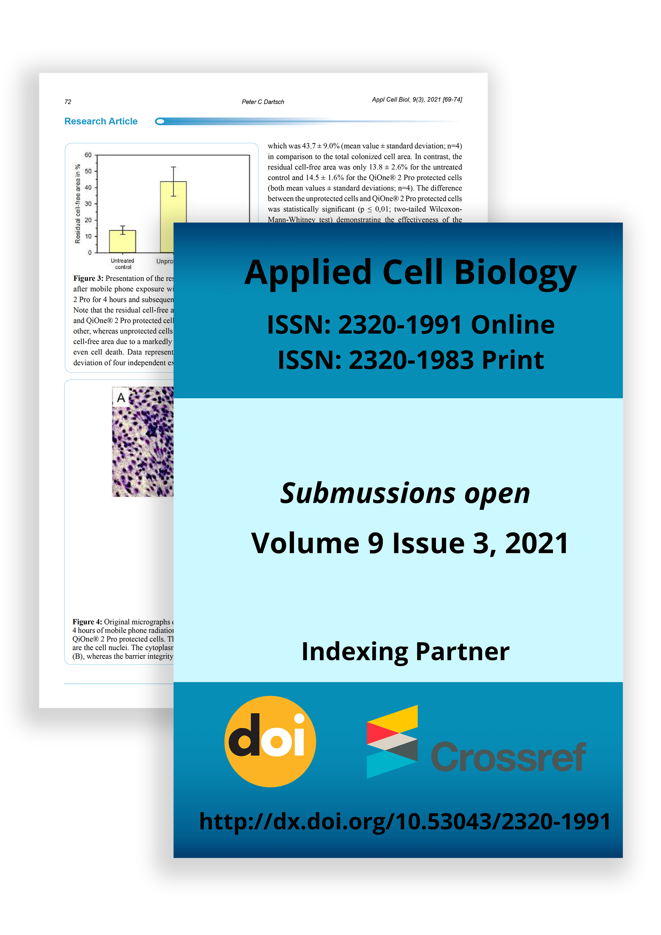
As seen in Figures 2 and 3, mobile phone radiation caused a reduction in cell regenerative activity by leaving a cell-free area which was 43.7 ± 9.0% (mean value ± standard deviation; n=4) in comparison to the total colonized cell area. In contrast, the residual cell-free area was only 13.8 ± 2.6% for the untreated control and 14.5 ± 1.6% for the QiOne® 2 Pro protected cells (both mean values ± standard deviations; n=4). The difference between the unprotected cells and QiOne® 2 Pro protected cells was statistically significant (p ≤ 0,01; two-tailed WilcoxonMann-Whitney test) demonstrating the effectiveness of the device against mobile phone radiation. Moreover, there was no statistically relevant difference between the QiOne® 2 Pro protected cells and the cells which were not exposed to mobile phone radiation at all.

Fig 3: Presentation of the results on cell regeneration after mobile phone exposure with and without QiOne® 2 Pro for 4 hours and subsequent cultivation for 8 hours. Note that the residual cell-free areas of untreated control and QiOne® 2 Pro protected cells do not differ from each other, whereas unprotected cells have a significant higher cell-free area due to a markedly reduced cell activity and even cell death. Data represent mean value ± standard deviation of four independent experiments.
Transepithelial Electrical Resistance (TEER)
As seen in Figure 4, mobile phone radiation caused a rupture on the epithelial barrier by causing cell death due to oxidative stress. In contrast, untreated controls and QiOne® 2 Pro protected cells did not show such massive morphological changes of the cell layers. This situation is reflected by the TEER values measured. For the unprotected cells a TEER of 152 ± 16 Ω/cm2 was measured demonstrating the complete loss of barrier integrity. The TEER value for the QiOne® 2 Pro protected cells was 1,837 ± 349 Ω/cm2 and for the untreated controls 2,542 ± 389 Ω/cm2 (all mean value ± standard deviation). The difference between protected and untreated cells was statistically not significant, whereas the difference to the unprotected cells was highly significant (p ≤ 0,01; twotailed Wilcoxon-Mann-Whitney test; n=3). When calculating the relative values by setting the TEER of the untreated controls as 100 ± 13.8%, the QiOne® 2 Pro protected cells had an value of 72.6 ± 14.5% and the unprotected cells of 6.0 ± 3.2% (all mean values ± standard deviations; Figure 5). Again, the data demonstrate the effectiveness of the device against mobile phone radiation.

Fig. 4: Original micrographs of IPEC-J2 cells establishing a physical barrier on the porous membranes of transwells after 4 hours of mobile phone radiation with subsequent cultivation for 24 hours. (A) Untreated control. (B) Unprotected cells. (C) QiOne® 2 Pro protected cells. The small dark points are the 0.4 µm pores in the membrane and the purple stained structures are the cell nuclei. The cytoplasm of the cells is only weekly stained by the dye. Note the rupture of the epithelial barrier in (B), whereas the barrier integrity in QiOne® 2 Pro protected cells in (C) is nearly similar to the untreated controls in (A).

Fig. 5: Presentation of the results on transepithelial electrical resistance (TEER) after mobile phone exposure with and without QiOne® 2 Pro for 4 hours and subsequent cultivation for 8 hours. Note that the TEER of untreated control and QiOne® 2 Pro protected cells do not differ from each other, whereas unprotected cells have a significant decreased TEER due to a rupture of the epithelial barrier. Data represent mean value ± standard deviation of three independent experiments.
4. Discussion
Bhattacharyya et al. [15] have summarized in their review that reactive oxygen species are generated as by-products of normal cellular metabolic activities which can be inactivated by endogenous enzymes such as superoxide dismutase, glutathione peroxidase and catalase. However, reactive oxygen species are generated as a response by a number of traumatic influences acting on our body. Among these are ultraviolet radiation, cigarette smoking, alcohol, nonsteroidal anti-inflammatory drugs, ischemia-reperfusion injury, chronic infections and inflammatory disorders.
Moreover, mobile phone radiation is also known to produce an excess of reactive oxygen radicals [10-13,16-20] which cannot be inactivated by the enzymes mentioned above. Since the epithelial cells of the intestinal barrier have a high turnover rate, they are more sensitive against oxidative stress which can induce oxidative injury and inflammatory responses involving a deficiency of the epithelium and immune/inflammation mediating cells [15].
Prompted by this background cultured intestinal cells were used to examine the effect of oxidative stress on the regenerative potential and the integrity of the epithelial barrier.
According to Vergauwen [21] “IPEC-J2 cells are intestinal porcine enterocytes isolated from the jejunum of a neonatal piglet. The IPEC-J2 cell line is unique as it is derived from the small intestine and is neither transformed nor tumorigenic in nature. IPEC-J2 cells mimic the human physiology more closely than any other cell line of non-human origin”. The cells were originally isolated in 1989 by Helen Berschneider at the University of North Carolina [22]. The advantage of the IPEC-J2 cell line as an in vitro model originates from its morphological and functional similarities with intestinal epithelial cells in vivo. IPEC-J2 cells have microvilli on their apical side and tight junctions to act as a barrier and reflecting epithelial functionality [23]. The determination of TEER is a technique that provides information about the uniformity of the IPEC-J2 cell layer on the microporous filter membrane and the integrity of the tight junctions formed between the polarized cells. Therefore, TEER measurements are often used to study epithelial barrier function [1].
The present results with QiOne® 2 Pro complement confirm our previous findings in which we demonstrated that this device was able to protect functional neutrophils against mobile phone radiation [24]. In these experiments, cell viability was checked by the generation of superoxide anion radicals in the course of an oxidative or respiratory burst. Mobile phone radiation in unprotected functional neutrophils caused a reduction in superoxide anion radical generation by approximately 40% in comparison to untreated cells. In contrast, QiOne® 2 Pro protected cells showed a reduction in superoxide anion radical generation by only 16% in comparison to untreated cells. In the present experiments with IPEC-J2 cells the regeneration values for unprotected and QiOne® 2 Pro protected cells were in the same range as in the previous experiments with functional neutrophils. However, the examination of the integrity of a three-dimensional intestinal barrier demonstrated a much higher sensitivity against oxidative stress from mobile phone radiation. This is in accordance to the findings reviewed by Bhattacharyya et al. [15]. The use of QiOne® 2 Pro protected the cells in a significant manner as shown here.
Although the principles of quantum electrodynamics (QED) are not really accepted in conventional medicine as a method to influence the state of water, the present investigation has shown that coherent water as generated by use of QiOne® 2 Pro obviously has a definite positive impact on cells by increasing their resistance against exogenous traumatic influences such as mobile phone radiation. It has been stated that electromagnetic fields can be coupled to coherent systems resulting in a “self-trapping” which causes a common in phase dynamical oscillation [25-28]. As a matter of fact, the coherent system might be protected against exogenous electromagnetic fields like the inner part of a Faraday cage.
HRV Measurements
Information regarding heart rate variability measurements can be found here:
https://www.defibrillator-deutschland.com – general information about HRV
http://edoc.sub.uni-hamburg.de – Master’s thesis in the medical technology course on HRV
A total of 7 HRV measurements were performed per subject. The following three measurements are shown graphically:
1st measurement: behavior of the body without additional stressor or Qi Blanco systems
2nd measurement: behavior of the body with an additional stressor
7th measurement: behavior of the body after 1 hour of exposure to the stressor; Qi Blanco systems are active
1. Rank Diagram
Stressor – inactive
Qi Blanco Systems – inactive
Stressor – active
Qi Blanco Systems – inactive
Stressor – active
Qi Blanco systems – active
2. Autonomous Nervous System [ANS] Status
Stressor – inactive
Qi Blanco systems – inactive
Stressor – active
Qi Blanco Systems – inactive
Stressor – active
Qi Blanco systems – active
3. Test Setup
3.1.1) Qi Blanco – QiOne endless
3.1.2) Qi Blanco – QiOne Master Prototyp
3.2 Measuring Equipment:
3.2.1) HF High Frequency Analyzer: 59B-HF Analyser Gigahertz Solutions – 27 MHZ – 25000 MHZ
Antenna: UBB27_G3 (Active, quasi-isotropic ultra-wideband antenna from 27 MHz to over 3.3 GHz)
3.2.2) HRV measuring device: HRV-Scanner standard Professional – Software Version 3.02.05
WLAN router: NETGEAR WG602 – 54 Mbps / IEEE 802.11b/g / 2.412 ~ 2.472 GHz
Radiation intensity: >20.00 µW/m²
Located at heart level of the test person at a distance of about 1 m.
Room size: approx. 16 m² – Room height 2.2 m
Basement, grass cover corresponds to ceiling height
Cellar window 1.0m x 0.5m closed – Ends approx. 0.2 m below the ceiling
House on corner property in quiet location with mostly older inhabitants, edge development, in the ground floor 3 visible WLAN networks.
In The Room Located Devices:
– 2 mobile phones – in flight mode
– 2 x laptops; 1x switched off, 1 x in flight mode to record HRV measurements
– 2 LED lamps hung centrally in the middle above the experimental setup (2x 64 Watt)
– Printer – switched off
– Stereo system – not connected
3.5 Electromagnetic Radiation During The Experiment:
1) Basic value: 3.90 – 3.98 µW/m²
2) Superimposed interference signal: every 22 sec a 4.2 sec long signal (origin unknown) 12.01 to >20.00 µW/m²
3.6 Measurement Procedure:
The HRV measurements were performed in a sitting position.
4. Experimental Procedure
The stressor was switched on from the 2nd measurement and used continuously until the 7th measurement was completed.
The Qi Blanco systems were used during the 3rd, 4th, 5th and 7th measurement.
Measurement, Date, Time, Duration, Rest Period, Stress Index, Median HF, Stressor active, QiBlanco System active, HRV-Age (short time age), Heartbeat per 5 min
5. Validation
The rest period between measurements was deliberately increased to emphasize the effect of the stressor and the Qi Blanco systems.
1) The low RF band function increases continuously over the test duration until it reaches a normal level of approx. 250 ms².
2) The deactivation of the Qi Blanco systems in test series 6 shows a jump in body activity in both cases.
3) The activation of the stressor is clearly visible in the records. The body is subject to high stress.
4) With active Qi Blanco systems, the body functions improve with increasing duration of exposure.
The validation is therefore successful.
6. Further Measurements
7. Note On The Tests
These are individual certificates. Every person is different and the respective effects are therefore individual. The tests presented here serve to qualitatively classify the effects of the Qi Blanco systems on the human body.
Literature
Detection of extraordinary large bio-magnetic field strength from human hand. Acupuncture and Electro-Therapuetics Research Interantional Journal 17:75-94
Sandyk R. 1995
Treatment of neurological and mental disorders. Patent No. 5, 470, 846
Jaffe L. F. 1981
The role of ionic currents in establishing developmental pattern. Philosophical Transactions of the Royal Society of London B 295:553-566]
Bassett C. A. L. 1995
Bioeletcromagnetics in the service of medicine. American Chemical Society, Washington DC
Oschman J. L. 2000
Energy Medicine. The Scientific Basis. – Churchill Livingstone
Become Part Of The Revolution!
- 100% Made In Germany
- Highest Quality Materials
- Worldwide Shipping